The effects of nanoparticles are being studied at the Faculty of Biology
-lg.jpg)
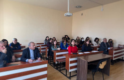
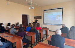
An academic seminar entitled “The Effect of Iron Nanoparticles on the Internal Organs and Embryonic Development Stages of Common Carp (Cyprinus carpio L., 1758)” was organized at the Faculty of Biology, delivered by Aysel Hajiyeva, a doctoral student of the Department of Biophysics and Biochemistry. The seminar was attended by the faculty administration, academic staff, and doctoral students.
Within the framework of the seminar, the results of the conducted research were presented. The study assessed the bioaccumulation properties of iron oxide nanoparticles (Fe₃O₄) in the small intestine and liver of common carp reared under aquaculture conditions, as well as their effects on embryonic development, using light and electron microscopy methods. The results demonstrated significant pathomorphological changes, including destruction of microvilli, cytoplasmic edema, and damage to mitochondria and vascular endothelium. It was determined that nanoparticles penetrated enterocytes, hepatocytes, and erythrocytes and accumulated systemically in various tissues.
At the same time, experiments on embryonic development showed that the addition of Fe₃O₄ nanoparticles at a dose of 0.001 g to sperm prior to fertilization increased the fertilization rate and the yield of healthy embryos by 12–14%. The presented results are significant for assessing the toxicity of nanoparticles at various stages of artificial fish reproduction and demonstrate potential applicability in aquaculture practice.
Seminar participants emphasized the scientific novelty and practical significance of the topic, noting the importance of more in-depth investigation of potential risks associated with nanoparticles, as well as their possible impacts on the environment and aquatic ecosystems.